Showing posts with label GIT. Show all posts

Possible factors for lower prevalence of GERD in Asia

Case History:
  • A 45 year old chronic  alcoholic and smoker comes to your clinic  with complaints of throat irritation and cough especially at night for 4 months duration.  The irritation is there in the morning but goes out as the day progresses. He has no other complaints except for occasional water brash.  His vitals are normal and physical examintaion reveals throat congestion. Rest of the examination is normal. How will you reach the diagnosis and manage this case?     
Gastroesophageal Reflux Disease(GERD)

Objectives
  1. Definition 
  2. Classification 
  3. Epidemiology 
  4. Etiology and Pathogenesis
  5. Clinical Manisfestations
  6. Diagnosis 
  7. Differential diagnosis 
  8. Treatment
1.Definition

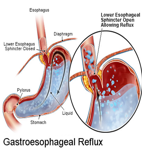
GERD is defined as chronic symptoms or mucosa damage produced by the abnormal reflux in the esophagus
Heartburn, water brash
superficial linear ulcer, erosion,redness,friability,bleeding,exudate,erythema,engorgement


2.  Classification

(1)  Non-erosive esophagitis (NERD)
(2)  Erosive esophagitis(EE) or reflux 
      esophagitis(RE) 
(3)  Barrett’s esophagus (BE)


(1)  Non-erosive esophagitis (NERD):
  •  the presence of typical symptoms of GERD caused by intra-esophageal acid
  •  the absence of visible esophageal mucosa injury at endoscopes

(2) Erosive esophagitis(EE) or reflux  esophagitis(RE) :
  •  the presence of typical symptoms visible esophageal mucosa injury at endoscopes

(3)Barrett’s Esophagus (BE)
  1. Acid damages lining of esophagus and causes chronic esophagitis
  2. Damaged area heals in a metaplastic process and abnormal columnar cells replace squamous cells

  • This specialized intestinal metaplasia(IM) can progress to dysplasia and adenocarcinoma
  • Many patients with Barrett’s are asymptomatic
3.  Epidemiology
  • GERD is one of the most prevalent gastrointestinal  disorders
  • GERD increases by age, the peak prevalence is in those people whose age is between 40-60ys.There is no difference between male and female
  • GERD in Asian population is not as usual in the Americans 
Possible factors for lower prevalence of GERD in Asia

(1)  Genetic factors
(2)  Low maximal acid output/small parietal cell  mass
(3) High LES(lower esophagus sphincter)  pressure
(4) Low BMI (body mass index)
(5) Lower consumption of alcohol, coffee, tea
(6) Lower or Less Smoking
(7) Fewer aggravating medicines
(8) Dietary factors(Lower dietary fat and chocolate in asian food)
(9) Lower consumption of carbonated soft drinks
(10)Lower consumption of citric fruit drinks

4.  Etiology and Pathogenesis

(1)Defense barrier against reflux consists of 

  1. tonic activity of the LES supported by crural diaphragm. 
  2. The ana’tomical location of the gastroesophageal junction below the diaphragmatic hiatus

  • Among them, the LES is primary barrier to gastroesophageal reflux , its basal  pressure is 10-30mmHg
  • LES normally works in conjunction with the diaphragm

  • If barriers disrupted, gastric contents go from stomach to esophagus.
  • In other words,reflux occurs only when the gradient of pressure between the LES and the stomach is lost.
  • The gradient of pressure lost  can be caused by a sustained or transient decrease in LES tone
A sustained hypotension of the LES (Defective basal LES pressure)may be due to :
  • muscle weaknes that is without apparent cause 
  • scleroderma-like disease, myopathy associated with chronic intestinal pseudo-obstruction, pregnancy, smoking, anticholinergic drugs, smooth-muscle relaxants[β-adrenergic drugs, aminophylline, nitrates, calcium channel blockers, phosphodiesterase inhibitors that increase cyclic AMP or cyclic GMP(including sildenafil), surgical damage to the LES, and esophagitis
  • Defective LES: transient LES relaxation (TLESR)increase frequency of reflux
  • TLESR without associated esophageal contraction is due to vagal reflex in which LES relaxation is elicited by gastric distention 
  • Increased episodes of TLESR are associated with GERD.
  • A similar reflex operates during belching.
Apart from incompetent barriers , reflux happens :
  1. when gastric volume is increased (after meal, in pyloric obstruction, in gastric stasis, during acid hypersecretion states)
  2. when gastric contents are near the gastroesophageal junction(in recumbency, bending down, hiatus hernia)
  3. when gastric pressure is increased (obesity, pregnancy, ascites, tight clothes)
(2) Esophageal clearance function
  •  It means that esophageal peristaltic contraction may clear materials refluxed to esophagus in normal condition
  • When Peristaltic contraction is impaired, Esophageal clearance is impaired
(3) Esophageal mucosal barrier
  •  It includes esophageal mucous epithelium and good blood circulation in submucous layer 
  •  Some factors ( e.g, smoking, alcohol, depression ) may injury this barrier and lead to RE.
(4) Esophageal mucosal injury produced by reflux
  • Exposure depends on the amount of refluxed material per episode, frequency of episodes, and rate of clearing the esophagus by gravity and peristaltic contractions
  • Acid refluxed into the esophagus is neutralized by saliva. Thus, impaired salivary secretion also increases esophageal exposure time
  • If the refluxed material extends to the cervical esophagus and breaches the upper sphincter , it can enter the pharynx, larynx and trachea

5. Clinical Manisfestations


(1)Esophageal symptoms --Most common symptoms

Heartburn—retrosternal burning discomfort
Regurgitation—effortless return of gastric contents into the pharynx without nausea, retching, or abdominal contractions
Dysphagia—difficulty(or trouble )swallowing
Chest pain 

(1)Esophageal symptoms -- Less common symptoms
  1. pain with swallowing (odynophagia)
  2. nausea

(2) Extraesophageal symptoms

Asthma, laryngitis, chronic cough, earache,  gingivitis, eroded tooth enamel, globus sensation, hoarseness, sore throat

(3) complications 
  1. Erosive esophagitis
  2. Peptic stricture
  3. Barrett’s esophagus
6. Diagnosis
  • If classic symptoms of heartburn and regurgitation exist in the absence of “alarm symptoms”,  the diagnosis of GERD can be made clinically and treatment can be initiated
(1) When to investigate?

Investigations are warranted if,
  a) the diagnosis is unclear
  b) symptoms persist or are refractory to treatment
  c) complications suspected with presence of alarm symptoms

※Alarm symptoms:
  1. BW loss > 10%
  2. Persistent vomiting
  3. Anemia, bleeding (Hematemesis, Melena)
  4. Progressive dysphagia or odynophagia
  5. Family UGI (upper gastrointestinal) cancer 

(2) Which investigation?

a) Barium swallow and meal: may be useful in: persistent dysphagia caused by suspected stricture or the assessment of large hiatus hernia
b) Endoscopes[highly specific (90-95%)] :
 (i) Most sensitive test for reflux esophagitis, diagnosis of other mucosal lesions
 (ii) Only sensitive method for diagnosing Barrette’s esophagus
 (iii) Recognition and management of peptic stricture

Trial of Medications
  • H2 blockers (H2 receptor blocking agents) or PPI (proton pump inhibitors)
  • Expect response in 2-4 weeks
  • If no response
  • Change from H2RA to PPI
  • Maximize dose of PPI


If PPI response inadequate despite maximal dosage 
  1. Confirm diagnosis
  2. EGD (Esophagogastrodudenoscopy)
  3. 24 hour pH monitoring 

Esophagogastrodudenoscopy (EGD)

Endoscopy (with biopsy if needed)

Patients who need EGD
  1. Alarm symptoms
  2. Poor therapeutic response
  3. Long symptom duration
  • Lacks sensitivity for identifying pathologic reflux
  • Absence of endoscopic features does not exclude a GERD diagnosis


7.Differential diagnosis
  • Symptoms of GERD may be similar to those of other diseases such as esophageal motility disorders, peptic ulcer,cholelithiasis, nonulcer dyspepsia, and angina pectoris
  • Furthermore, RE may be confused with pill-induced damage, radiation esophagitis ,or infections (cytomegalo’virus, herpes, Candida)

8. Treatment

Goals of therapy
  • Symptomatic relief
  • Heal esophagitis
  • Avoid complications
Current Management for GERD

(1) Life-style modification
(2) Medical therapy 
(4) Surgery
(5) Management of Complications

(1) Lifestyle modifications
  • Avoid large meals (Avoid over food)
  • Avoid acidic foods (citrus/tomato), alcohol, caffiene, chocolate, onions, garlic, peppermint
  • Decrease fat intake
  • Avoid lying down within 3-4 hours after a meal
  • Elevate head of bed 4-8 inches – very effective
  • Avoid meds that may potentiate GERD (CCB, alpha agonists, theophylline, nitrates, sedatives, NSAIDS)
  • Avoid clothing that is tight around the waist
  • Lose weight
  • Stop smoking

(2) Medical therapy for GERD
  • Antacids
  • Proton Pump Inhibitors(PPI)
  • H2 blockers
  • Prokinetic agents: Cisapride, Metoclopramide, Erythromycin

(4) Antireflux surgery
  • Antireflux surgery( indications)
  • Failed medical management
  • Patient preference
  • GERD complications
  • Medical complications attributable to a large hiatal hernia
  • Atypical symptoms with reflux documented on 24-hour pH monitoring

(5) Management of Complications

Barrett’s Esophagus
  • Manage in same manner as GERD
  • EGD every 3 years in patients without dysplasia
  • In patients with dysplasia annual to shorter interval surveillance

(5) Management of Complications
  • Esophageal stricture
  • May need dilation

Distinguishing characteristics of CD and UC

Case History :
  • A 5 year old boy is brought to your clinic with complaints of several episodes of diarrhoea each day for 2 years duration. There is also complaint of failure to gain weight properly for the same duration. He appears ill. He has pallor and dehydration. On examination, his pulse is 150 bpm and his BP is 95/40mmHg. He is mildly febrile. You also notice some depigmented macules on his forearm and legs. He is at 5th percentile for weight and 60th percentile for height. What is your provisional diagnosis and how will you manage this case?
  • A 30 year old male comes to your clinic with complaints of crampy abdominal pain and bloody diarrhea for past 10 days.  He also complains of mild intermittent fever for the same duration.  In the past medical history, he tells you that he has been having several similar episodes for past 5 years. He occasionally takes over the counter medicines sometimes for his illness otherwise it goes away on its own most of the time. He also gives you a history of occasional knee and ankle joints pain for past 2 years. On examination, the patient is thin built. He appears dehydrated and mildly anemic. There is no abdominal tenderness. Rest of the examination is normal. How will you manage this case?

Inflammatory Bowel Disease (IBD) is a
general term given to two diseases:
- Ulcerative colitis
- Crohn's disease

IBD characterized by a tendency for chronic or relapsing immune activation and inflammation within the GIT.
Crohn’s disease (CD) and ulcerative colitis (UC) are the 2 major forms of idiopathic IBD.

LESS COMMON ENTITIES OF IBD ARE:

Microscopic colitis (collagenous and lynphocytic)
Others
  • Diversion colitis
  • Radiation colitis
  • Drug induced colitis
  • Infectious colitis
  • Ischemic colitis 

CD is a condition of chronic inflammation potentially involving any location of the GIT from mouth to anus.

UC is an inflammatory disorder that affects the rectum and extends proximally to affect variable extent of the colon.

Genetics

Studies suggested that 1st degree relatives of an affected patient have a risk of IBD that is 4-20 times higher than that of general population.
The best replicated linkage region, IBD1, on chromosome 16q contains the CD susceptibility gene, NOD2/CARD15.
Having one copy of the risk alleles confers a 2–4-fold risk for developing CD, whereas double-dose carriage increases the risk 20–40-fold.

Epidemiologic Features of Inflammmatory Bowel Disease

Incidence (per100,000) 1-10(CD), 2-18 (UC)
Prevalence (per100,000) 20-100 (CD), 40-100 (UC)
Race White >black> Hispanic
Sex M=F
Age at Onset Peak  15-25, second peak 50- 80(CD)
Smoking Associated with CD,inversely associated with UC
Relapse Nonsteroidal anti-inflammatory drugs , oral contraceptives (CD)

  • Both illnesses do have one strong feature in common. 
  • They are marked by an abnormal response of the body's immune system.
The symptoms of these two disorders can be very similar, but there are also two distinct differences:
  • the part of the intestinal tract affected
  • the extent (severity) of the inflammation

Etiology
  • Mutations within the NOD2/ CARD15 gene contribute to CD susceptibility.
  • Functional studies suggest that inappropriate responses to bacterial components may alter signaling pathways of the innate immune system, leading to 
  • the development and persistence of intestinal inflammation.
  • Initiating pathogen?
  • Infectious?
  • ? Possibly non-pathogenic commensal enteric flora

UC: PATHOLOGY
  • The inflammation is predominantly confined to the mucosa.
  • Non-specific (can be seen with any acute inflammation)
  • The lamina propria becomes edematous.
  • Inflammatory infiltrate of neutrophils
  • Neutrophils invade crypts, causing cryptitis & ultimately crypt abscesses.
  • Specific (suggest chronicity):
  • Distorted crypt architecture, crypt atrophy and a chronic inflammatory infiltrate.


Distinguishing characteristics of CD and UC
Endoscopic features of CD and UC
Pathologic features of CD and UC


How IBD is Diagnosed

Clinical history
  • Physical examination
  • Laboratory tests
  • Endoscopy (Gastroscopy/Colonoscopy)
  • X-ray (contrast) findings 
  • Tissue biopsy (pathology)
  • Exclude other possibilities (need good history, physical exam, labs, imaging and endoscopy with biopsy)
  • There are many distinguishing features of CD and UC.
  • In about 5% it is classified as indeterminate because of overlapping features.
Inflammatory Bowel Disease:

Symptoms
The most common symptoms of IBD are:
  1. abdominal pain
  2. diarrhea
  3. Rectal bleeding
  4. weight loss and fever may also occur
  5. vomiting, and loss of appetite
  6. skin and eye irritations
  • Inflammation invariably involves the rectum. ( sigmoid and whole)  
  • The lesions in ulcerative colitis tend to occur in continuous regions of the gut
  •  The inflammation affects mainly the inner lining of the bowel (the mucosa)
  • Inflammation is confluent and is more severe distally. In long standing pancolitis  the bowel  becomes shortened and ‘pseudopolyps’ develop
  • Histologically  the inflammaory process is limited to the mucosa and spares the deeper layers of the bowel wall.
  • Both acute and chronic inflammaory cells infiltrate  te lamina propria and the crypts. Crypt abscesses are typical.Goblet cells lose their mucus and in long standing cases glands become distorted.
Symptoms of Ulcerative Colitis
  • Symptoms depend on extent and severity of inflammation
  • Rectal bleeding and urgency to evacuate
  • Diarrhea
  • Abdominal cramping
–Extraintestinal (systemic) symptoms
  • Joint pain/swelling
  • Eye inflammation
  • Skin lesions
Physical Signs
  • No specific clinical signs on general examination
  • Tenderness at left iliac fossa
  • Acute illness may reveal peritoneal irritation findings
  • Rectal examination may reveal blood and mucus on the glove
In adults at presentation about 55% have proctitis, 30% left sided colitis and 15% extensive colitis or pan colitis

  • Rectum is almost always involved
  • The diseased areas are contiguous
  • Histology reveals crypt abscess and inflammatory polyps


Laboratory
  • Leucocytosis
  • Anemia
  • ESR elevation
  • Hypoalbuminemia
  • Electrolyte imbalance


Radiology
Double contrast barium enema exam of the colon
(lead pipe appearance)

  • Associated Autoimmune Diseases
  • Pernicious anemia
  • Systemic lupus erythematosus
  • Hashimoto’s thyroiditis

IBD : Management Goals of Crohn's Disease and Ulcerative colitis

    Crohn’s disease 
    1. Crohn’s disease can affect any part of the digestive tract
    2. Crohn’s disease lesions are often discontinuous
    3. Affects all layers of the bowel from the inner mucosa to the outer serosa
    • The sites most commonly involved, in order of frequency are terminal ileum, and right side of colon, colon alone, terminal ileum alone, ileum and jejunum.
    • Characteristically, the entire wall of the bowel is oedematous and thickened.
    • There are deep ulcers which appear as linear fissures; thus the mucosa between them is described as cobblestone 
    • Deep ulcers  may penetrate  through  the bowel wall to initiate abscesses or fistulae. Fistulae  may develop between adjacent loops of bowel or between affected segments of bowel and the bladder, uterus or vagina and may appear perineum. 
    • Characteristically  the changes  are patchy . Even when short segment of bowel is affected  the inflammatory process is interrupted by islands of normal mucosa and the changes from the affected part is abrupt. 
    • Skip area.
    • The mesenteric lymph nodes are enlarged and the mesentery thickened.
    • Histologically, chronic inflammation is seen through all the layers of the bowel wall which is thickened as a result. There are focal  aggregates or microgranuloma are seen and when they are near to the surface of the mucosa they often ulcerate  to form tiny aphthous –like ulcers.
    Common Symptoms of Crohn’s Disease
    • Diarrhea
    • Abdominal pain and tenderness
    • Loss of appetite and weight
    • Fever
    • Fatigue
    • Rectal bleeding (less as compared to UC) and anal ulcers
    • Stunted growth in children


    CROHN’S DISEASE

    Macroscopically, thickening due to edema and fibrosis, which can be severe enough to cause a stricture
    Granuloma formation on the serosal surface
    “Cobblestone” appearance due to a combination submucosal edema and fissuring ulcers
    Apthoid (mild superficial) and fissure (severe deep) ulcers


    Clinical Features

    Crohn’s disease 

    Presentation depends o the major site of disease involvement
    Ileal- abdominal pain associated with diarrhoea which is watery 

    No blood or mucus
            
    Weight loss 
            
    Features of nutritional deficiency

    • Rectal sparing and the presence of perianal disease are features which favour a diagnosis of Crohn’s disease rather than ulcerative colitis.
    • Features of both small bowel and colonic disease.
    • Physical exam-weight loss, anaemia with glossitis, angular stomatitis,  abdominal tenderness and abdominal mass


    Smoking in IBD
    • Ulcerative Colitis
    • Smoking can protect against UC
    •  Ex-smokers are more likely to develop UC


    Crohn’s disease
    • Twofold risk in current smokers
    • Smokers are less responsive to treatment
    • Recurrence of disease after surgery


    Complications

    Intestinal 
    •  Severe life-thratenening inflammation of the colon
    •  Perforation of the small intestine  or colon
    •  Life threatening acute hemorrhage
    • Fistula and perianal disease


    Extra intestinal
    • Ocular
    • Liver
    • Mesenteric or portal vein thrombosis
    • Venous thrombosis
    • Arthralgia of large joints
    • Erythema nodosum
    • Pyoderma gangrenosum 
    • Metabolic bone disease


    Nutritional complications are common in IBD:

    deficiencies of proteins, calories, and vitamins.
    These deficiencies may be caused by inadequate dietary intake, or poor absorption (malabsorption).
    Other complications associated with IBD include:
      arthritis, skin rashes, arthritis, inflammation of the eye or mouth, kidney stones, or other diseases of the liver and biliary system.


    Treatment

    • no cure for IBD but there is medical therapy and surgical therapy
    •    Medical therapy for IBD has three main goals:
    •  Inducing remission (periods of time that are
    •     symptom-free)
    • Maintaining remission (preventing flare-ups
    •    of disease)
    • Improving the patient’s quality of life

    At the present time, there are five basic categories of medications used in the treatment of IBD. They are:
    • Aminosalicylates
    • Corticosteroids
    • Immunomodulators
    • Antibiotics
    • Biologic therapies

    5-Aminosalicylic Acids
    • The mainstay treatment of mild to moderately active UC and CD (induction).
    • 5-ASA may act by 
    • blocking the production of prostaglandins and leukotrienes,
    • inhibiting bacterial peptide–induced neutrophil chemotaxis and adenosine-induced secretion, 
    • scavenging reactive oxygen metabolites
    • For patients with distal colonic disease, a suppository or enema form will be most appropriate.
    • Maintenance treatment with a 5-aminosalicylic acid can be effective for sustaining remission in ulcerative colitis but is of questionable value in Crohn's disease. 

    Corticosteroids
    • Topical corticosteroids can be used as an alternative to 5-ASA in ulcerative proctitis or distal UC. 
    • Oral prednisone or prednisolone is used for moderately severe UC or CD, in doses ranging up to 60 mg per day. 
    • IV is warranted for patients who are sufficiently ill to require hospitalization; the majority will have a response within 7 to 10 days.
    • No proven maintenance benefit in the treatment of either UC or CD. 
    • Many and serious side effects. 
    • Budesonide: 
    • less side effects, 
    • its use is limited to patients with distal ileal and right-sided colonic disease

    Immunosuppressive Agents
    • These agents are generally appropriate for patients in whom the dose of corticosteroids cannot be tapered or discontinued. 
    • Azathioprine & 6-MP 
    • The most extensively used immunosuppressive agents.
    • The mechanisms of action unknown but may include 
    • suppressing the generation of a specific subgroup of T cells through inhibiton of calcineurin. 
    • The onset of benefit takes several weeks up to six months.
    • Dose-related BM suppression is uniformly observed
    • Methotrexate 
    • Effective in steroid-dependent active CD and in maintaining remission.
    • Cyclosporine 
    • Severe UC not responding to IV steroid &needing urgent proctocolectomy.
    • 50% of the responders will need surgery within a year.
    • TNF inhibitors
    • Infliximab
    • Etanercept


    INDICATIONS FOR SURGERY

    In patients with UC:
    • Severe attacks that fail to respond to medical therapy. 
    • Complications of a severe attack (e.g., perforation, acute dilatation). 
    • Chronic continuous disease with an impaired quality of life. 
    • Dysplasia or carcinoma.
    In patients with CD
    • Obstruction, severe perianal disease unresponsive to medical therapy, difficult fistulas, major bleeding, severe disability
    • 30 % relapse rate
    IBD Sequelae 

    UC:
    • Risk of cancer begins after 8 years, risk of pancolitis 7% at 20 years and 17% at 30 years.
    • Increased risk for cancer: early age of onset, pancolitis.
    • Need for colonoscopic screening after 8 years
    CD:
    • True incidence of cancer is uncertain, but could be as high as UC
    • Need the same screening policy.

    What does the general physician need to know about Malabsorption ?


    Malabsorption syndrome 

    Malabsorption is a broad term used to describe the inability to absorb nutrients through the gut lining into the bloodstream. This means the failure of the GI tract, usually the small intestine, to absorb one or more substances from the diet. This is generally the result of some defect or damage to the mucosal lining of the small intestine where most of our nutrient absorption takes place. 

    • Malabsorption is not a disease by itself, but rather the result of some other condition that is present. 
    • Malabsorption may affect one or more of the many nutrients present in the diet, including protein, fat, carbohydrate, vitamins, and minerals. 

    • Celiac disease (sprue, gluten - sensitive enteropathy) 
    • Crohn's disease 
    • The degree of malabsorption depends on the type of underlying condition and the extent to which it has affected the gut. Some of the more common malabsorption syndromes are due to bacterial or parasitic infections, 

    • Tropical sprue
    •  Liver disease (including cirrhosis, hepatitis, and gallstones),
    • Whipples disease 
    • Lactase deficiency 
    • Parasitic diseases 
    • Other causes of malabsorption can be due to past intestinal surgeries, bacterial overgrowth, AIDS, radiation to the abdomen, diabetes, lymphoma or motility disorders 
    The most common symptoms of malabsorption include:
    • Diarrhea, steatorrhea (excessive amount of fat in the stool), and abdominal distention with cramps, bloating, and gas due to impaired water and carbohydrate absorption, and irritation from unabsorbed fatty acids. The individual may also report explosive diarrhea with greasy, foul-smelling stools. 

    • Anemia, with weakness and fatigue due to inadequate absorption of vitamin B12, iron, and folic acid 

    • Edema (fluid retention in the body's tissues) due to decreased protein absorption 
    • Malnutrition and weight loss due to decreased fat, carbohydrate, and protein absorption. Weight may be 80-90% of usual weight despite increased oral intake of nutrients. 
    • Muscle cramping due to decreased vitamin D, calcium, and potassium levels 
    • Muscle wasting and atrophy due to decreased protein absorption and metabolism 
    • Perianal skin burning, itching, or soreness due to frequent loose stools.


    Risk factors for malabsorption syndrome include
    • premature birth 
    • family history of malabsorption or cystic fibrosis 
    • use of certain drugs, such as mineral oil or other laxatives 
    • travel to foreign countries 
    • intestinal surgery, including bowel transplantation 
    • excess alcohol consumption.
    Celiac disease (sprue, gluten - sensitive enteropathy) is a condition where the mucosal lining of the small intestine is damaged by ingestion of gluten. Gluten is a protein found in wheat, rye, barley and oats but not in corn or rice. The mechanisms and reason for this reaction are unknown, but it is thought to be genetic in nature and likely an autoimmune condition and is not considered an allergy. 

    Maintaining a gluten-free diet restores the damaged mucosal lining to normal and allows one to absorb normally leading to a normal life. On occasion, patients may fail to respond to a gluten-free diet and could have refractory sprue or intestinal lymphoma.


    Tropical sprue 
    • is a malabsorptive disorder more often in people from the Caribbean, India, or southeast Asia and should be suspected in anyone presenting with malabsorption that has visited the tropics.
    •  Although its cause is unknown, it is thought to be related to environmental factors, including infection, intestinal parasites, or possibly the consumption of certain food toxins.
    •  Symptoms often include a sore tongue, anemia, weight loss, along with diarrhea and passage of fatty stools. Treatment consists of antibiotics, often Tetracycline, for up to 6 months. 

    Whipples disease 
    • is a systemic bacterial illness, Tropheryma whipplei,  usually affecting middle age. 
    • The cause is thought to be related to bacterial infection, resulting in nutritional deficiencies, chronic low-grade fever, diarrhea, joint pain, weight loss, and darkening of the skin's pigmentation. 
    • Other organs of the body may be affected, including the brain, heart, lungs, and eyes. 
    • It is diagnosed by taking a small bowel biopsy through an endoscope, and the treatment is antibiotics for one year or longer 

    Lactase deficiency 
    • can result in malabsorption and occurs in 15 percent of Caucasian, 50 percent of blacks and nearly 90 percent of Asians
    • Often times, however, one can have a partial lactase deficiency that causes symptoms but not full-blown malabsorption. Treatment is to avoid lactose-containing dairy products (milk, ice cream ) 

    Short bowel syndromes 
    • may be present at birth (congenital) or the result of surgery-reduce the surface area of the bowel available to absorb nutrients and can also result in malabsorption syndrome. 
    • Congenital short bowel syndrome occurs in about 24 out of 100,000 live births and has a high mortality rate (about 38%).

    Diagnosis of malabsorption
    • The diagnosis of malabsorption syndrome and identification of the underlying cause can require extensive diagnostic testing. 
    • First phase - a thorough medical history and physical examination by a physician, who will then determine the appropriate laboratory studies and radiological studies to assist in diagnosis.
    • A 72-hour stool collection may be ordered for fecal fat measurement; increased fecal fat in the stool collected indicates malabsorption. 
    • A biopsy of the small intestine
    •  Ultrasound, computed tomography scan (CT scan), magnetic resonance imaging (MRI), barium enema, or other x rays to identify abnormalities of the gastrointestinal tract and pancreas may also be ordered.
    Investigations for malabsorption and its causes

    Routine – 

    • stool microscopy and its culture
    • Fe, folate, Vit. B12
    • Ca, albumin, alk. Phosphatase, Mg,
    • Celiac antibodies 


    Specific-

    • H2 breath testing – sugar
    • Fecal elastase
    • Duodenal biopsy
    • Barium studies
    • D-xylose  test
    • Schilling test
    • SeHCAT test


    Treatment
    • Fluid and nutrient monitoring and replacement is essential for any individual with malabsorption syndrome. 
    • Hospitalization may be required when severe fluid and electrolyte imbalances occur. 
    • Consultation with a dietitian to assist with nutritional support and meal planning is helpful. If the patient is able to eat, the diet and supplements should provide bulk and be rich in carbohydrates, proteins, fats, minerals, and vitamins. The patient should be encouraged to eat several small, frequent meals throughout the day, avoiding fluids and foods that promote diarrhea. 
    • Intake and output should be monitored, along with the number, color, and consistency of stools.
    • The individual with malabsorption syndrome must be monitored for dehydration, including dry tongue, mouth and skin; increased thirst; low, concentrated urine output; or feeling weak or dizzy when standing.
    •  Pulse and blood pressure should be monitored, observing for increased or irregular pulse rate, or hypotension (low blood pressure). 
    • The individual should also be alert for signs of nutrient, vitamin, and mineral depletion, including nausea or vomiting; fissures at corner of mouth; fatigue or weakness; dry, pluckable hair; easy bruising; tingling in fingers or toes; and numbness or burning sensation in legs or feet.
    •  Fluid volume excess, as a result of diminished protein stores, may require fluid intake restrictions. The physician should also be notified of any shortness of breath. 
    Specific medical management  dependents upon the cause.
    • Tropical sprue consists of folic acid supplements and long-term antibiotics
    • Whipple's disease also may require long-term use of antibiotics, such as tetracycline.
    • Injections of vitamin B12 in terminal ileal disease.
    • Enzymes to replace missing intestinal enzymes, or antispasmodics to reduce abdominal cramping and associated diarrhea
    Prognosis
    • The expected course for the individual with malabsorption syndrome varies depending on the cause. 
    • The onset of symptoms may be slow and difficult to diagnose. 
    • Treatment may be long, complicated, and changed often for optimal effectiveness. 
    • Patience and a positive attitude are important in controlling or curing the disorder. 
    • Careful monitoring is necessary to prevent additional illnesses caused by nutritional deficiencies.

    How will you proceed with management of pancreatitis?

    Case History:

    A 45 year old lady comes to your clinic with complaints of severe pain in epigastric region for 6 hours duration. The pain is sharp stabbing type with radiation to the back. There is slight relief of her pain on leaning forward. She also has vomited twice since her pain began. She gives you a history of incidental diagnosis of cholelithiasis two years back. On examination, she looks very distressed. Her pulse is 100bpm, BP is 100/60mmHg, RR is 24/min. There is significant tenderness in epigastric region with rigidity and guarding. What is your provisional diagnosis? How will you proceed with management of this case?  

    Pancreatitis

    Pancreatic inflammatory disease may be

        classified as (1) acute pancreatitis and (2) chronic pancreatitis.

    Acute Pancreatitis

    The pathologic spectrum of acute pancreatitis varies from edematous pancreatitis, which is usually a mild and self-limited disorder, to life threatening necrotizing pancreatitis.
    The incidence of pancreatitis varies in different countries and depends on cause, e.g., alcohol, gallstones, metabolic factors, and drugs.

    Causes of Ac. Pancreatitis

    Common Causes
    • Gallstones (including microlithiasis): 30-60%
    • Alcohol (acute and chronic alcoholism): 15-30%
    • Hypertriglyceridemia
    • Endoscopic retrograde cholangiopancreatography (ERCP), especially after biliary manometry
    • Trauma (especially blunt abdominal trauma)
    • Postoperative (abdominal and nonabdominal operations)
    • Drugs (azathioprine, 6-mercaptopurine, sulfonamides, estrogens, tetracycline, valproic acid, anti-HIV medications)
    • Sphincter of Oddi dysfunction
    Uncommon causes
    • Vascular causes and vasculitis (ischemic-hypoperfusion states after cardiac surgery)
    • Connective tissue disorders and thrombotic thrombocytopenic purpura (TTP)
    • Cancer of the pancreas
    • Hypercalcemia
    • Periampullary diverticulum
    • Pancreas divisum
    • Cystic fibrosis
    • Renal failure
    Rare causes
    • Infections (mumps, coxsackievirus, cytomegalovirus, echovirus)
    • Autoimmune (e.g., Sjo¨gren’s syndrome)

    Causes to consider in patients with recurrent bouts of acute pancreatitis without an obvious etiology
    • Occult disease of the biliary tree or pancreatic ducts, especially microlithiasis, sludge
    • Drugs
    • Hypertriglyceridemia
    • Pancreas divisum
    • Pancreatic cancer
    • Sphincter of Oddi dysfunction
    • Cystic fibrosis
    • Idiopathic

    Pathogenesis
    • Acute pancreatitis occurs as a consequence of premature activation of zymogen granules,  releasing proteases which digest the pancreas and surrounding tissue 
    • The severity of acute pancreatitis is dependent upon the balance between activity of released proteolytic enzymes and antiproteolytic factors. The latter comprise an intracellular pancreatic trypsin inhibitor protein and circulating β2-macroglobulin, α1-antitrypsin and Cl-esterase inhibitors.
    • Usually mild and self-limiting with minimal organ involvement however in some cases, it is severe with local complications such as necrosis, pseudocyst or abscess, and systemic complications leading to multi-organ failure. 
    • The mortality in this severe group is approximately 30%. 


    Clinical Features

    Symptoms
    • upper abdominal pain and the pain usually begins in the epigastrium accompanied by nausea and vomiting.
    •  As inflammation spreads throughout the peritoneal cavity the pain becomes more intense.
    • There is radiation of the pain to the back with relief on bending forward because of involvement of the retro-peritoneum. 
    Signs
    • Only upper abdominal tenderness in mild cases
    • In more severe disease, the patient may have a tachycardia, hypotension and be oliguric.
    • Abdominal examination may show widespread tenderness with guarding as well as reduced or absent bowel sounds.
    • Specific clinical signs that support a diagnosis of severe necrotizing pancreatitis include periumbilical (Cullen's sign) and flank bruising (Grey Turner's sign).


    Investigations
    • Serum amylase – within 24 hours, the rise is   > 3 times normal, the level falls down over next 3-5 days; persistent elevation is suggestive of pseudocyst formation
    • Serum lipase – also raises in early period but its level remains elevated for 7 – 15 days, so important in cases of late presentation
    • Other baseline investigations
    • a full blood count , CRP, urea and electrolytes, blood glucose, liver biochemistry, plasma calcium and arterial blood gases
    • These are documented at presentation and then repeated at 24 and 48 hours and provide a basis for assessing the severity of an attack and prognosis.


    Radiological investigations
    • Erect chest X-ray- to exclude gastro-duodenal perforation which may also raise amylase
    • Abdominal USG- to exclude cholelithiasis and choledocholithiasis
    • Contrast-enhanced spiral CT scanning - essential in all except for mild attacks of pancreatitis. Carried out within 2-3 days of presentation, it allows the extent of pancreatic necrosis to be assessed.
    • MRI or MRCP – particularly useful in differentiating solid and fluid inflammatory masses


    Assessment of severity

    Factors during the first 48 hours that indicate severe pancreatitis and a poor prognosis (three or more factors present predict a severe episode)
    • Age  >55 yrs
    • WBC  > 15,000/cmm
    • Blood glucose >10mmol/l
    • Serum urea  >16mmol/l
    • Serum albumin <30g/dl
    • Serum aminotransferase >200U/l 
    • Serum calcium <2mmol/l
    • Serum LDH >600U/l
    • PaO2 <60mmHg
    In addition, serial assessment of C-reactive protein (CRP) is a useful indicator of progress. A peak CRP > 210 mg/l in the first 4 days predicts severe acute pancreatitis with 80% accuracy.

    Complications of Acute Pancreatitis

    LOCAL
    • Necrosis
    • Sterile
    • Infected
    • Pancreatic fluid collections
    • Pancreatic abscess
    • Pancreatic pseudocyst
    • Pain
    • Rupture
    • Hemorrhage
    • Infection
    • Obstruction of gastrointestinal tract (stomach, duodenum, colon)
    • Pancreatic ascites
    • Disruption of main pancreatic duct
    • Leaking pseudocyst
    • Involvement of contiguous organs by necrotizing pancreatitis
    • Massive intraperitoneal hemorrhage
    • Thrombosis of blood vessels (splenic vein, portal vein)
    • Bowel infarction
    • Obstructive jaundice

    SYSTEMIC

    Pulmonary
    • Pleural effusion
    • Atelectasis
    • Mediastinal abscess
    • Pneumonitis
    • Adult respiratory distress syndrome
    Cardiovascular
    • Hypotension
    • Hypovolemia
    • Sudden death
    • Nonspecific ST-T changes in electrocardiogram simulating myocardial infarction
    • Pericardial effusion
    Hematologic
    • Disseminated intravascular coagulation
    • Gastrointestinal hemorrhage
    • Peptic ulcer disease
    • Erosive gastritis
    • Hemorrhagic pancreatic necrosis with erosion into major blood vessels
    • Portal vein thrombosis, variceal hemorrhage

    Renal
    • Oliguria
    • Azotemia
    • Renal artery and/or renal vein thrombosis
    • Acute tubular necrosis
    Metabolic
    • Hyperglycemia
    • Hypertriglyceridemia
    • Hypocalcemia
    • Encephalopathy
    Central nervous system
    • Psychosis
    • Fat emboli
    • Fat necrosis
      Treatment
      • Early fluid losses in acute pancreatitis may be large, requiring well-maintained intravenous access as well as a central line and urinary catheter to monitor circulating volume and renal function.
      • Nasogastric suction: to prevent aspiration pneumonia
      • Baseline arterial blood gases determine the need for continuous oxygen administration.
      • Prophylactic antibiotics: cefuroxime or aztreonam
      • Analgesia requirements. Pethidine and tramadol are the drugs of choice. The morphine derivatives should be avoided because they can cause sphincter of Oddi contraction.
      • Feeding. In patients with a severe episode there is little likelihood of oral nutrition for a number of weeks. Total parenteral nutrition has been associated with a high risk of infection and has been replaced by enteral nutrition. This is administered via a nasojejunal tube, which is well tolerated and can maintain adequate nutritional input.
      • In a small proportion of patients, multiorgan failure will develop in the first few days after presentation reflecting the extent of pancreatic necrosis. Such patients will require positive-pressure ventilation and often renal support. 
      • These patients may also need surgical debridement of necrotic pancreatic tissue.
      • The mortality in this group is extremely high (in excess of 80%).

      Treatment of gallstone related pancreatitis
      • In patients with gallstone-related pancreatitis and associated cholangitis, endoscopic intervention with sphincterotomy and stone extraction is of proven benefit and is the treatment of choice. 
      • In the absence of cholangitis, sphincterotomy and stone extraction is only of proven benefit when the episode of pancreatitis is predicted as severe.
      • In less severe cases of gallstone-related pancreatitis intervention for gallstone can be deferred until full recovery is obtained (an approximate 6-week period).


      Prognosis

      Mortality: 10-15%
      One-third occur within the first week, usually from multi-organ failure. After this time the majority of deaths result from sepsis, especially that complicating infected necrosis.

      Pathophysiology of Chronic Pancreatitis

      • Chronic pancreatitis is a chronic inflammatory disease characterised by fibrosis and destruction of exocrine pancreatic tissue.
      • Diabetes mellitus occurs in advanced cases because the islets of Langerhans are involved. 

      Pathophysiology
      • Around 80% of cases in Western countries result from alcohol misuse 
      • In developing countries malnutrition and associated dietary factors have been implicated.


      Other causes
      • Cystic Fibrosis
      • Benign and malignant processes obstructing pancreatic duct
      • Congenital anomaly like pancreatic divisum
      • Rarely, inherited as autosomal dominant with variable penetrance


      Clinical features 

      • Chronic pancreatitis predominantly affects middle-aged alcoholic men
      • Pain is the most common presentation of chronic pancreatitis and is usually epigastric and often radiating through into the back.
      • In 50%,episodes of 'acute pancreatitis‘ with each attack resulting in a degree of permanent pancreatic damage.
      • In 35%, relentless, slowly progressive chronic pain without acute exacerbations
      • remainder have no pain but diarrhoea
      • Exocrine and endocrine insufficiency may develop at any time, and occasionally malabsorption or diabetes are the presenting features in the absence of abdominal pain.
      • Steatorrhoea
      • Protein malabsorption
      • Diabetes Mellitus
      • Weight loss is common and results from a combination of anorexia, avoidance of food because of post-prandial pain, malabsorption and/or diabetes


      Complications

      Pseudocyst - < 6cm, usually resolves spontaneously; > 6cm, requires intervention
        Ascites
      Pleural effusion
      Strictures – CBD, duodenum
      Portal hypertention


      Investigations

      The extent dependens upon the clinical setting.
      In a patient with known alcohol abuse and typical pain few confirmatory tests are required.

      Tests performed include:
      • Faecal elastase level will be abnormal in the majority
      • Serum amylase and lipase levels are rarely significantly elevated in established chronic pancreatitis
      • Pancreolauryl test
      • Transabdominal ultrasound scan is used for initial assessment.
      • Contrast-enhanced spiral CT scan – test of choice
      • MRCP
      • Endoscopic ultrasound – specifically for assessing complications
      • Diagnostic ERCP – replaced by MRCP in developed countries


      CT scan showing a grossly dilated and irregular duct with a calcified stone (arrow A). Note the calcification in the head of the gland (arrow B). 


      MRCP of the same patient showing marked ductal dilatation with abnormal dilated side branches (arrows A). A small cyst is also present (arrow B).


      Endoscopic retrograde cholangiogram shows grossly dilated pancreatic ducts (arrows) in a patient with long-standing pancreatitis

      Treatment
      • Alcohol avoidance is crucial in halting the progression of the disease and reducing pain.
      Pain relief 
      • Analgesics particularly NSAIDs are useful
      • Opiates reserved for chronic unremitting pain and often leads to addiction
      • Oral pancreatic enzyme supplementation reduces pancreatic secretion of enzymes and thus provides pain relief in some patients.
      • Coeliac plexus neurolysis or minimally invasive thoracoscopic splanchnicectomy – provides long term pain relief but pain eventually recurs

      Patients who are abstinent from alcohol and who have severe chronic pain which is resistant to conservative measures are considered for intervention with following procedures:
      •  Endoscopic therapy
      Dilatation or stenting of pancreatic duct strictures 
      Removal of calculi (mechanical or shock-wave lithotripsy) 
      Surgical methods
      Partial pancreatic resection, preserving the duodenum 
      Pancreatico-jejunostomy 
      Total pancreatectomy – high morbidity and mortality with the procedure

      • Steatorrhoea - dietary fat restriction , oral pancreatic enzyme supplements; a proton pump inhibitor is added to optimise duodenal pH for pancreatic enzyme activity
      • Management of complications